Vertebral Compression Fracture Case Study
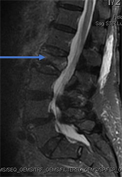
88 year old female fell in the bathroom with increasing amount of back pain. Despite pain pills in the hospital, she could not get out of bed. MRI showed compression fracture due to osteoporosis (weak bone). MRI can show if the fracture is new based on swelling in the bone. The arrow shows the bone is whiter than the other bones indicating a new fracture.
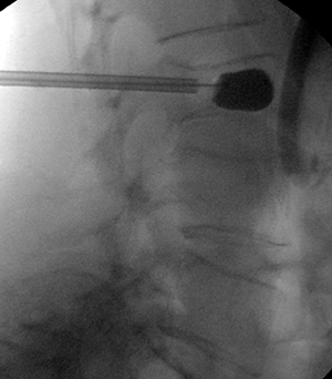
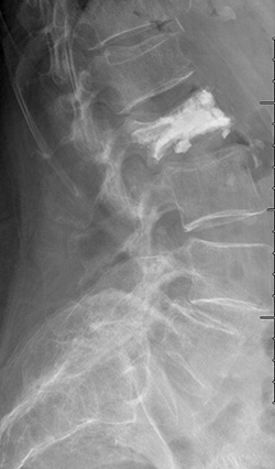
She underwent minimally invasive kyphoplasty to treat the fracture. Through a pen hole incision a balloon is placed inside the bone to restore the bone back to normal shape as possible. (It never looks quite normal). Then cement is placed into the bone and acts like grout making the fracture all healed.


